
Researchers use lab-grown tissue grafts for personalized joint replacement
most recent study, published today inScience Translational Medicine, builds upon a long series of their previous developments that began in 2005 on bioengineering functional cartilage and bone for regenerative medicine and tissue models of disease.
The authors used the Yucatan minipig to establish their methodology for TMJ reconstruction using the recipients’ own cells. The team isolated the stem cells from a small amount of fat obtained from each animal, expanded the cells in culture to obtain a sufficient number for a large graft, and induced them into the cartilage and bone- forming cells.
Using imaging-guided fabrication, the researchers shaped a block of clinically used decellularized bovine bone matrix into the exact geometry of the TMJ being repaired.
They infused this scaffold with bone-forming cells, while inducing cartilage formation by compacting a 1-mm thick surface layer of condensed mesenchymal cells.
They built the matching bioreactor chamber so that the scaffold fitted tightly into it, like a hand in a glove.
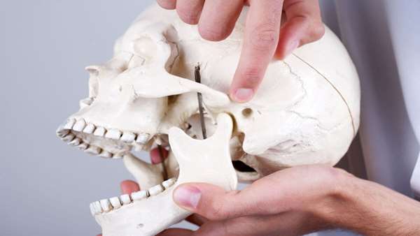
The temporomandibular joint (TMJ), which forms the back portion of the lower
jaw and connects your jaw to your skull, is an anatomically complex and highly loaded structure
consisting of cartilage and bone. About 10 million people in the United States alone suffer from TMJ
dysfunction due to birth defects, trauma, or disease. Current treatments range from steroid
injections that provide only a temporary pain relief, to surgical reconstructions using either prosthetic
devices or donor tissue, and often fail to provide long-lasting repair. Researchers have sought a
better way to treat TMJ, including investigating biological TMJ grafts grown in the lab that could
integrate with the native tissues, remodel the joint over time, and provide life-long function for the
patient.
Reference: https://medicalxpress.com/pdf521882010.pdf
